LS2 Live

Double illumination, double detection light sheet microscope for optimal volumetric imaging of multiple large samples.

LS2 Live

Double illumination, double detection light sheet microscope for optimal volumetric imaging of multiple large samples

Long-term live imaging

Controlled temperature and gas concentrations enable culturing and imaging of large 3D samples in physiological conditions.

Easy sample mounting

Open-top configuration allows easy sample mounting and medium exchange even during running time lapse.

Multiplex-imaging experiments

Multi-position imaging with the possibility to enable independent imaging parameters for each imaged position.

Dedicated multi-well sample holder

  • Samples are held in sample chamber of custom shapes adaptable to the sample being imaged.

  • Easy sample mounting from top and medium exchange possible during running time-lapse
.
  • Sample can be placed in different media (e.g drugs vs control experiments).

Double illumination

  • Two 10X objectives are used to illuminated from below.

  • Light sheet generated by scanning gaussian beams (DLSM) of tunable thickness.

  • Illumination lasers across all visible range from (405nm to 680nm).

Dual view detection

  • Collection of the emitted light from two sides for optimal volumetric imaging.

  • Choice of high NA objectives (25X 1.1 NA or 16x 0.8 NA)
.
  • Possibility to collect transmitted light images of the sample.

Flexible microscope software

User friendly and intuitive instrument operation design makes light sheet imaging very accessible.

Do you need to delete a position, extend a Z range or change laser intensity without stopping a running time-lapse? It’s possible!

Intelligent imaging: automatically adapt to the live image acquired by the system and adjust measurement parameters.

Technical specifications

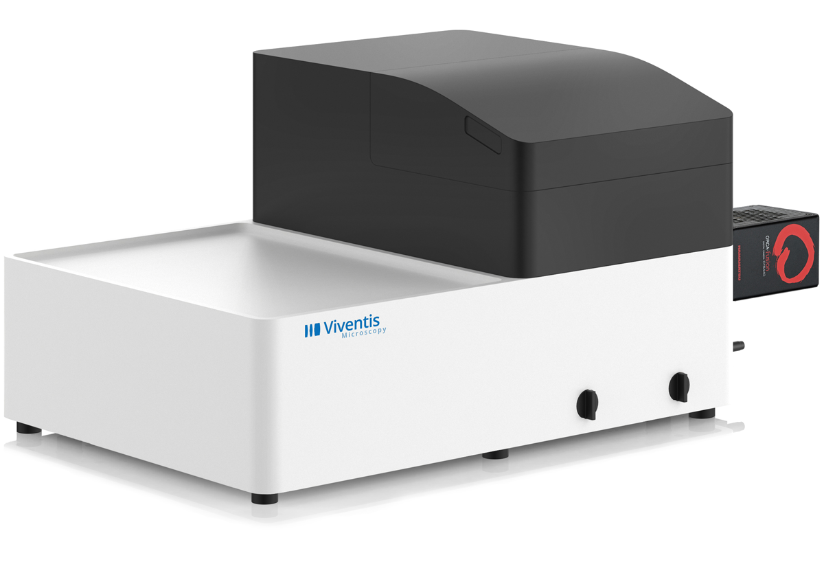
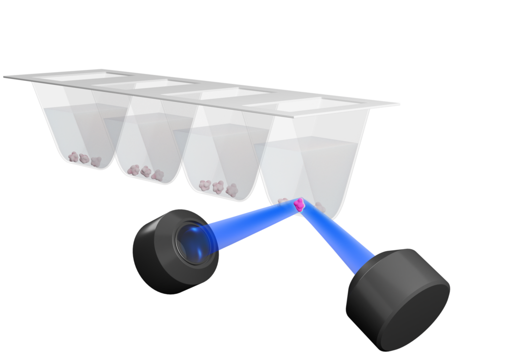
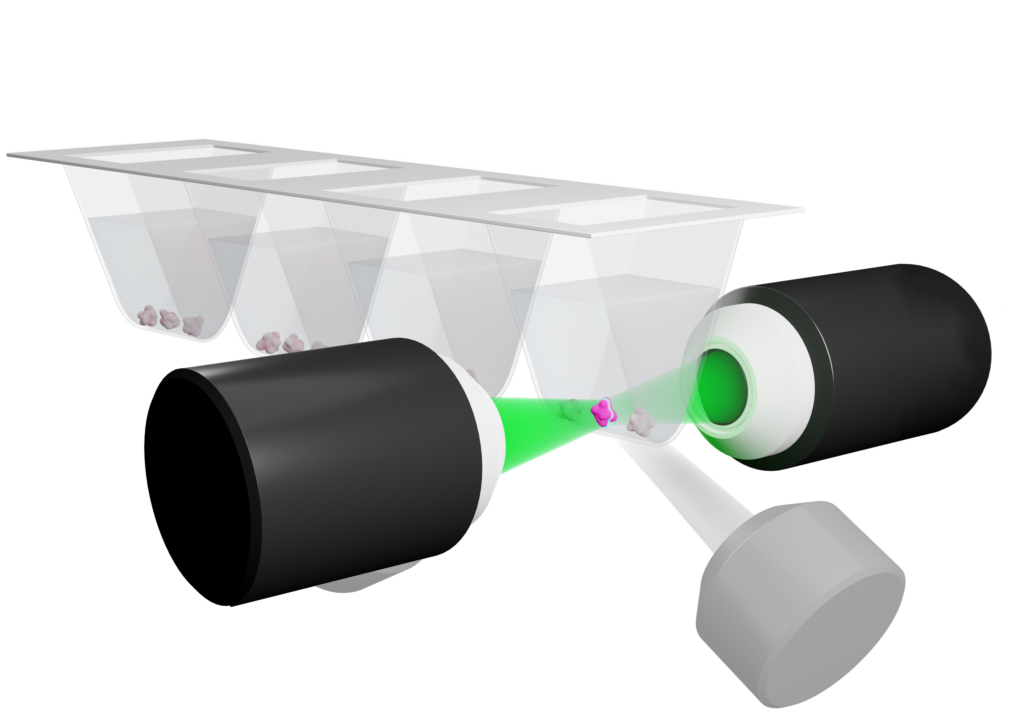
Detection and Illumination optics

  • Illumination: two Nikon 10X objectives, NA 0.2.

  • Detection: two Nikon 25X NA 1.1 OR two Nikon 16X NA 0.8 water immersion objectives.

Illumination

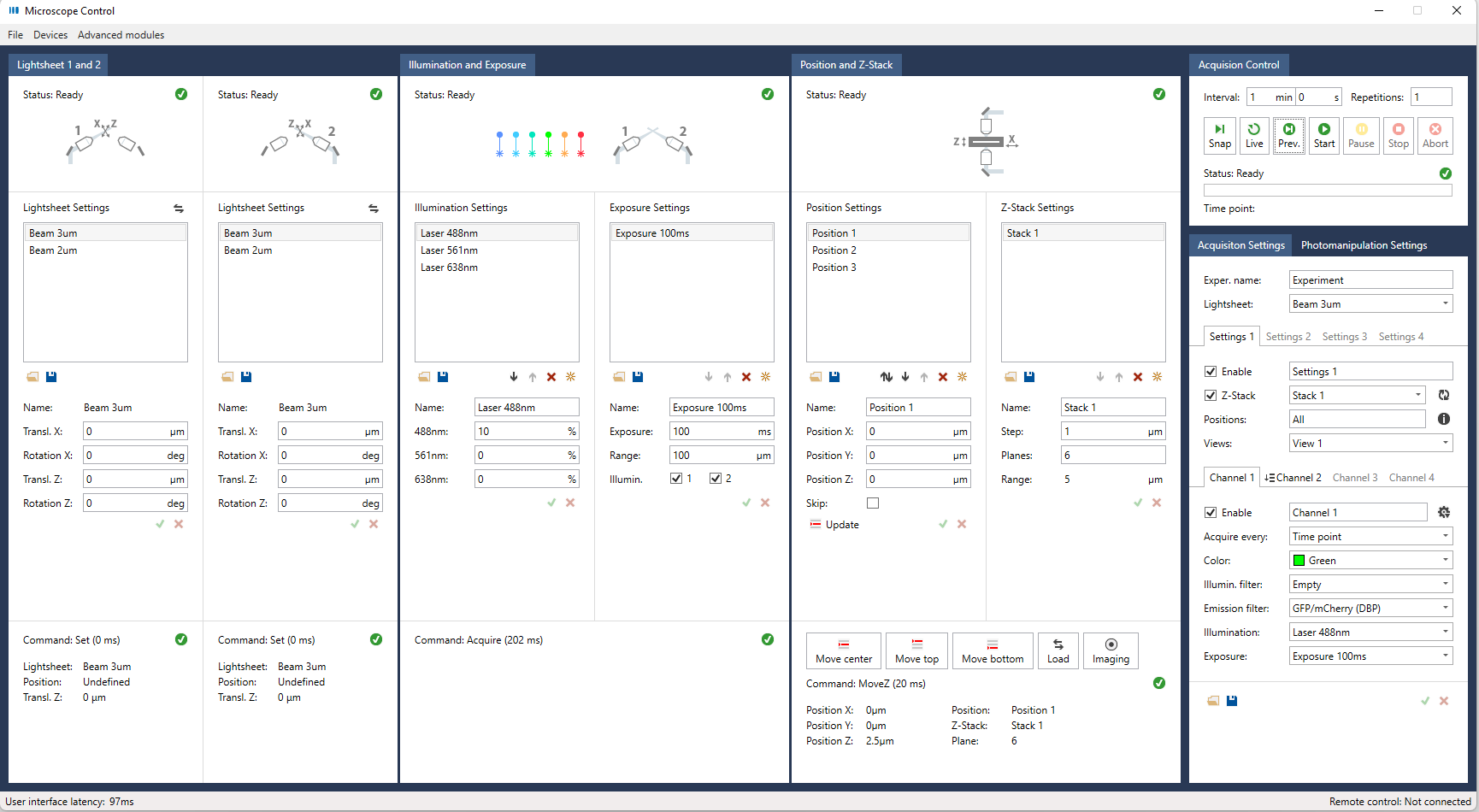
  • External laser combiner with a maximum of six wavelengths (from 405nm to 685nm), Omicron LightHUB+.

  • Transmitted light illumination (LED light source) to locate the sample and acquire transmitted images.

Light sheet specification

  • Light sheet generated by scanning of a Gaussian beam.

  • Three switchable light-sheets with thicknesses (FWHM) of approximately 2.1, 3.5 and 4.5 μm for different sample sizes.

  • Automatic position-specific light-sheet alignment.

Detection camera

  • Two high sensitivity Hamamatsu ORCA-Fusion USB3 cameras.

  • Field of view: 16X Objective: 900µm; 25X Objective: 596µm

  • Pixel size: 16X Objective: 406nm; 25X Objective: 260nm
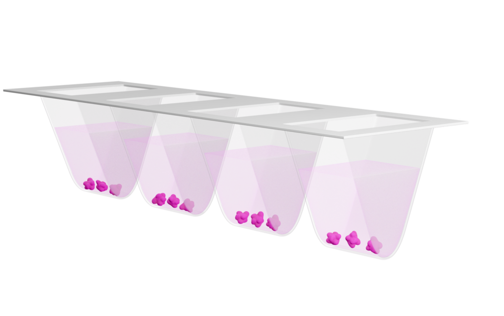
Sample part

  • Motorized XYZ sample stage with maximum X travel range of 50mm

  • Multiple sample located in a open-top FEP sample chamber (multi-well).

Sample incubation

  • Recirculating air temperature controller from 5°C above ambient temperature to 40°C.

  • CO2 concentration control range 4-10%.

  • O2 concentration control range 5-15%.

Imaging Applications

Explore the wide range of imaging of application where our light-sheet systems have made an impact on the research of many scientists.

Imaging Applications

Explore the wide range of imaging of application where our light-sheet systems have made an impact on the research of many scientists.

Useful links

Viventis Microscopy Sàrl
EPFL Innovation Park, Building A
1015 Lausanne, Switzerland

info@viventis-microscopy.com