LS1 Live

High resolution live imaging based on double illumination and single detection light sheet microscopy.

Long-term live imaging

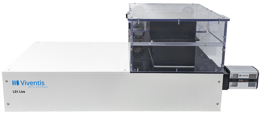
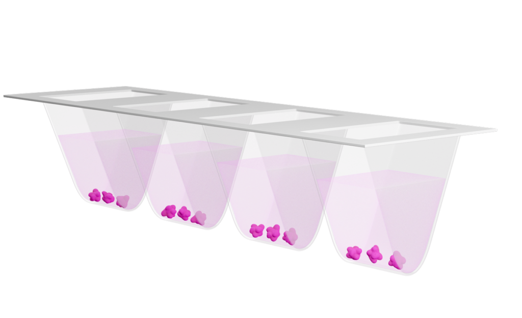
Controlled temperature and gas concentrations enable culturing and imaging of large 3D samples in physiological conditions.

Easy sample mounting

Open-top configuration allows easy sample mounting and medium exchange even during running time lapse.

Multiplex-imaging experiments

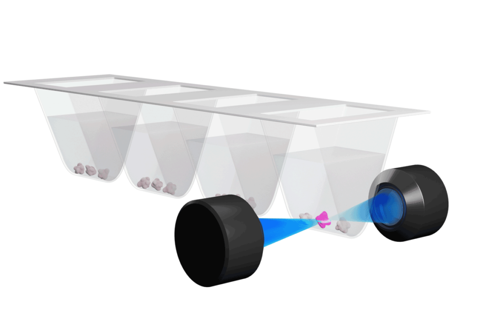
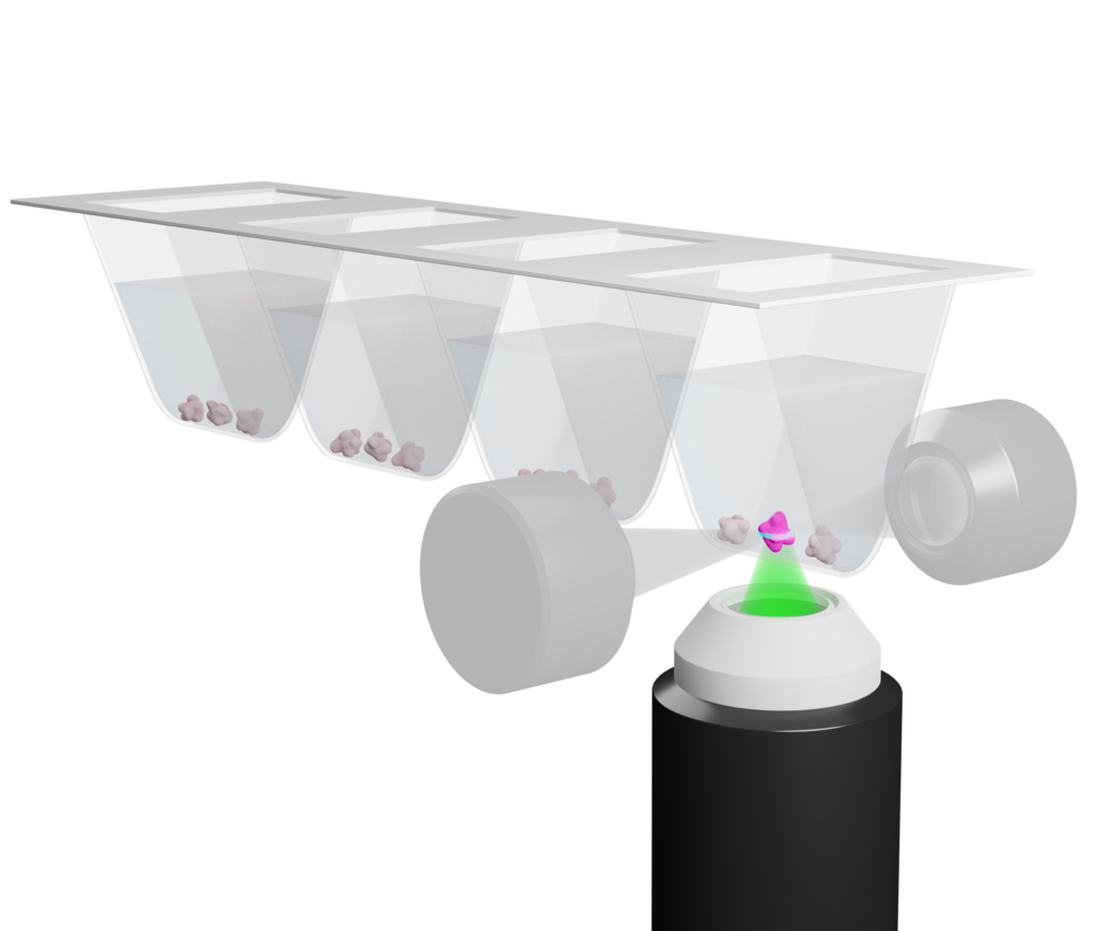
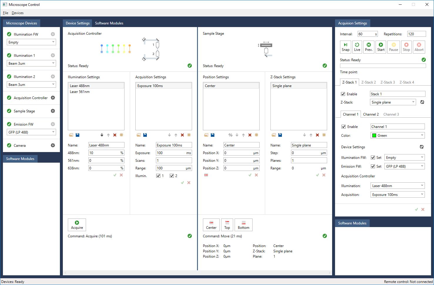
Multi-positions imaging with the possibility to enable independent imaging parameters for each imaged position.

Dedicated multi-well sample holder

  • Samples are held in sample chamber of custom shapes adaptable to the sample being imaged.

  • Easy sample mounting from top and medium exchange possible during running time-lapse
.
  • Sample can be placed in different media (e.g drugs vs control experiments).

Double illumination

  • Two opposing 10X objectives are used to illuminate the sample from the side.
  • Light sheet generated by scanning gaussian beams (DLSM) of tunable thickness.

  • Illumination lasers across all visible range from (405nm to 680nm).

Single view detection

  • 25X 1.1 NA objective for optimal sub-cellular resolution imaging.
  • Switchable magnification for a field of view from 400µm to 800µm.
  • Possibility to collect transmitted light images of the sample.


Flexible microscope software

User friendly and intuitive instrument operation design makes light sheet imaging very accessible.

Do you need to delete a position, extend a Z range or change laser intensity without stopping a running time-lapse? It’s possible!

Intelligent imaging: automatically adapt to the live image acquired by the system and adjust measurement parameters.

Technical specifications

Detection and Illumination optics

  • Illumination: two Nikon 10X objectives, NA 0.3.

  • Detection: one Nikon 25X NA 1.1 water immersion objective with switchable magnification (18x and 37X).

Illumination

  • External laser combiner with a maximum of six wavelengths (from 405nm to 685nm), Omicron LightHUB+.

  • Transmitted light illumination (LED light source) to locate the sample and acquire transmitted images.

Light sheet specification

  • Light sheet generated by scanning of a Gaussian beam.

  • Three switchable light-sheet with thicknesses (FWHM) of approximately 1.5, 2.2 and 3.3 μm for different sample sizes.

  • Automatic position-specific light-sheet alignment.

Detection camera

  • One high sensitivity Hamamatsu ORCA-Fusion USB3 cameras.

  • Field of view: 18X: 800µm; 37X: 400µm

  • Pixel size: 18X 347nm; 37X 173nm

Sample part

  • Motorized XYZ sample stage with maximum X travel range of 50mm

  • Multiple sample located in a open-top FEP sample chamber (multi-well).

Sample incubation

  • Recirculating air temperature controller from 5°C above ambient temperature to 40°C.

  • CO2 concentration control range 4-10%.

  • O2 concentration control range 5-15%.

Imaging Applications

Our light-sheet systems have made a positive impact on the reserach of many scientists. Explore the wide-range of imaging application.

Imaging Applications

Explore the wide range of imaging of application where our light-sheet systems have made an impact on the research of many scientists.

Useful links

Viventis Microscopy Sàrl
EPFL Innovation Park, Building A
1015 Lausanne, Switzerland

info@viventis-microscopy.com